|
|
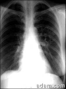
This chest X-ray shows the affects of the fungal infection, coccidioidomycosis. In the middle of the left lung (seen on the right side of the picture) there are multiple, thin-walled cavities (seen as light areas) with a diameter of 2 to 4 centimeters. To the side of these light areas are patchy light areas with irregular and poorly defined borders. Diseases that may explain these X-ray findings include lung abscesses, chronic pulmonary coccidioidomycosis, chronic pulmonary tuberculosis, chronic pulmonary histoplasmosis, and others.
|